Case Report - (2021) Volume 7, Issue 7
Lipoma arborescens is a rare benign type of synovial membrane tumor. It occurs due to the replacement of synovial tissue by mature adipocytes. It usually affects the knee and is monoarticular. Despite a typical imaging examination, the final diagnosis is only possible with histopathological examination of the lesion. The objective of this article is to present a case diagnosed in a tertiary hospital in Porto Alegre and to highlight this pathology as a differential diagnosis for oncological knee injuries. An 18-year-old female patient had an enlarged left knee for 7 years. We opted for open resection of the lesion with wide synovectomy. One year later, the patient showed improvement in clinical complaints and no signs of tumor recurrence. Although rare, lipoma arborescens should be considered in the differential diagnosis of knee pathologies and open synovectomy is currently the treatment of choice.
Lipoma arborescens, Knee tumor, Synovial tumor
Lipoma arborescens is a rare benign type of synovial membrane tumor. It occurs due to replacement of synovial tissue by mature adipocytes. In most cases, it is monoarticular and affects the knee. Clinically, it presents as an increase in the volume of the affected joint. Magnetic resonance imaging usually presents a typical pattern, able to differentiate it from other pathologies of the knee. However, the final diagnosis is only possible with histopathological examination of the lesion. The aim of this article is to present a case diagnosed in a tertiary hospital in Porto Alegre and highlight this pathology as a differential diagnosis for knee injuries.
An 18-year-old female patient started outpatient care at a university hospital in Porto Alegre complaining of a mass in her left knee. She reported a 7-year history of pain and progressive joint growth for 5 years. On physical examination, she presented a significant increase in volume in the anterior region of the left knee, which was softened, painless and immobile. She had an incompetent extensor mechanism. On the radiographs of the left knee, he presented involvement of the lower pole of the patella, with extensive calcification, without the presence of other bone lesions.
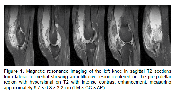
Magnetic resonance imaging of the left knee (Figure 1) showed, in the extensor compartment, extensive joint effusion in greater volume in the suprapatellar recess, diffusely thickened joint synovium, digitiform images with a sign of fat within the joint space and significant edema of the Hoffa’s fat. She presented an infiltrative lesion centered on the prepatellar region with hypersignal on T2 and hyposignal on T1, with intense contrast enhancement, measuring approximately 6.7 × 6.3 × 2.2 cm (lateromedial, cephalocaudal and anteroposterior dimensions). The lesion also affected the patella, which was reduced, with destruction of the cortical and bone marrow. The patellar tendon was partially disrupted and there was tendinosis of the quadriceps tendon. These findings could indicate villonodular synovitis, giant cell tumor and arborescent lipoma.

Figure 1: Magnetic resonance imaging of the left knee in sagittal T2 sections from lateral to medial showing an infiltrative lesion centered on the pre-patellar region with hypersignal on T2 with intense contrast enhancement, measuring approximately 6.7 × 6.3 × 2.2 cm (LM × CC × AP).
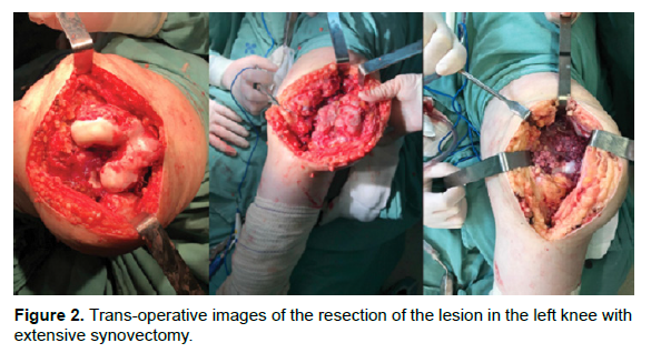
Chest computed tomography staging did not show secondary implants. The patient underwent a biopsy with an inconclusive result, showing only the presence of chronic osteomyelitis in the left patella. An open resection of the lesion was then indicated, with extensive anterior synovectomy, respecting the medial and lateral insertion areas and preserving the meniscal area (Figure 2). The femoral, patellar and tibial chondral matrix was also regularized, and the extensor component was retensioned. Patella stability was tested at the end of surgery, with no signs of instability. Anatomopathological examination of the synovial tissue and soft parts of the left knee showed chronic synovitis with suppurative areas, synovial hyperplasia, granulation tissue and reparative fibrosis.

Figure 2: Trans-operative images of the resection of the lesion in the left knee with extensive synovectomy.
Twelve months after resection, he was undergoing physiotherapy and presented, on physical examination, a reduction in the volume of the left knee compared to the physical examination prior to surgery, although it was still increased in relation to the contralateral side. He had a competent extensor mechanism and left knee flexionextension of 10 to 100°. Magnetic resonance imaging of the left knee did not show tumor recurrence.
Lipoma arborescens is an uncommon type of benign synovial fluid tumor. Its most common location is on the knee, followed by the shoulder, elbow, wrist, hip and ankle [1]. It can affect some extraarticular structures such as tendons and bursae [2]. In most cases, it is unilateral; when they are bilateral or affect different joints, they are considered atypical cases. It affects men and women in the same proportion, usually between the fourth and fifth decades of life. The youngest patient reported was 9 years old and the oldest 90 years old [3].
Its etiology and pathophysiology are unknown, although many cases are related to previous trauma, diabetes, steroid use, or degenerative or inflammatory arthritis. It is characterized by a replacement of synovial tissue with mature adipocytes. The clinical features of these tumors are generally similar to other conditions of the knee, characterized by a progressive and chronic increase in the volume of the joint without pain, associated with joint effusion. With the progression of the disease, it leads to restriction of movement of the joint [2,3].
Magnetic resonance imaging (MRI) is the exam of choice [4]. It presents as a villous mass arising from the synovia with an isointense fat signal [5]. Differential diagnosis should be made with free bone bodies, synovial chondromatosis, “rice bodies” (present in rheumatoid arthritis), pigmented villonodular synovitis, synovial hemangioma, intra-articular lipoma and intra-articular liposarcoma, among others [6,7].
Other diagnostic tests include laboratory tests, synovial fluid aspirate, and other imaging tests. Laboratory tests are useful to exclude systemic diseases such as rheumatoid arthritis. Synovial fluid aspirate is primarily used to exclude other causes of swelling and joint effusion. On radiographs, an increase in soft tissue density, bone erosion, subchondral sclerosis, osteophytes, among other findings of osteoarthritis, or no changes may appear. On ultrasound, it is possible to visualize the villi projected into the joint. Computed tomography helps to exclude other differential diagnoses [4,6].
Macroscopically, it has a proliferating yellowish appearance with villous projections arranged in an arborescence pattern, usually occupying the suprapatellar pouch. Histologically, the villi are filled with mature fat cells. As imaging findings are highly suggestive, some authors report that biopsy is not necessary prior to treatment [1,4].
The treatment of choice is open synovectomy. Recurrence after surgery is not common. Video-arthroscopy and chemical synovectomy have been reported, but there were more cases of recurrence. Evolution to malignancy has not yet been described for this type of tumor. Patients in whom treatment with complete resection is not performed early are more likely to develop osteoarthritis of the affected joint [1,5].
In this case, followed up by our service, the patient presented a clinical picture similar to reports in the literature. Open resection of the lesion was chosen to relieve the patient's complaints and exclude the possibility of any malignancy. Although rare, lipoma arborescens should be considered in the differential diagnosis of knee pathologies, and open synovectomy is the most used treatment so far, with good results, and, therefore, it is currently the treatment of choice.
We thank the patient for allowing the case description.
No funding was needed for the case description.
The authors declare that they have no conflicts of interest.
Citation: Daniela B, et al. Lipoma Arborescens – A Case Report. Oncol Cancer Case Rep. 2021, 07 (7), 001-002.
Received: 09-Jun-2021 Published: 22-Jul-2021
Copyright: © 2021 Daniela B, et al. This is an open-access article distributed under the terms of the Creative Commons Attribution License, which permits unrestricted use, distribution, and reproduction in any medium, provided the original author and source are credited.