Case Report - (2019) Volume 11, Issue 1
Meckel’s diverticulum is a rare gastrointestinal congenital anomaly that is difficult to diagnose and presents with anemia, abdominal pain, or intussusception was described originally by Guilhelmus Fabricius Hildanus in 1598. [1,2] A Meckel’s diverticulum is usually an asymptomatic condition, and many cases are incidentally discovered during a radiographic evaluation or during surgery performed for other reasons. [1] The diagnosis is usually made in childhood. Here, we report the case of a 44-year-old female with a volvulated Meckel’s diverticulum, causing a small bowel obstruction. The patient initiated with profuse diarrhea and vomiting bilious content; then started with abdominal distention and severe abdominal pain, becoming an intestinal obstruction. Later, the patient was surgically intervened; finding a Meckel’s diverticulum volvulated in the intraoperative that was causing the obstruction.
Meckel's diverticulum is rarely diagnosed in adults. It is usually asymptomatic and becomes evident when complicated. [3] Of the few reported cases; intestinal obstruction is the most common presentation in adult and is the second most common in children. [3]
Meckel’s diverticulum, Volvulus, Intestinal obstruction, Abdominal pain
Meckel diverticulum is the most frequent congenital anomaly of the gastrointestinal tract, occurring in 2% to 3% of the population, as a consequence of the failure in the obliteration of the vitelline duct during embryological development. [3,4] The prevalence reported between men and women is 2:1. [2] The diagnosis is usually made in childhood; between 50% and 60% of patients which develop symptoms are under 2 years old. 2 As the complications associated with Meckel's diverticulum tend to decrease with increasing age, Meckel's diverticulum is rarely diagnosed in adults. On average, the diverticulum is located two feet proximal to the ileocecal valve and often contains gastric or pancreatic tissue, causing mucosal bleeding. [5] The symptomatology is secondary to diverticulum complications such as ulceration and hemorrhage; diverticulitis; intestinal obstruction due to diverticular inversion, intussusception, volvulus, torsion or inclusion of the diverticulum in a hernia. [2]
A 44-year-old female; with no significant past medical history; presented to the emergency room complaining of liquid, yellow and foul-smelling stools in 20 occasions; accompanied by diffuse, colicky abdominal pain, with intensity 8/10 on the pain scale; followed by vomiting of gastric contents in 3 occasions and general weakness. In the emergency was treated with antiemetics and hydration without improvement. Physical examination showed a slightly distended abdomen with hyperactive bowel sounds, painful to deep palpation and no masses or hernias were identified. The patient is admitted with a diagnosis of Acute Gastroenteritis. Laboratory tests were normal, with a negative stool test, without ova or parasite. Plain abdominal X-ray demonstrated an undetermined pattern of intestinal air distribution, with discrete gas in rectal ampulla and air fluid levels can be seen at the midline and left hypochondrium.
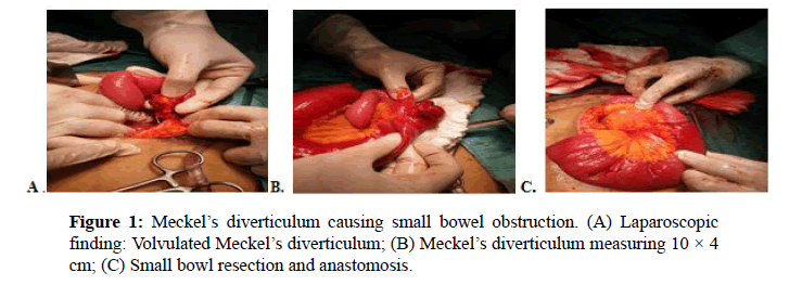
On the following day, the patient presented febrile peaks (38.5 C) and continued with severe abdominal pain, vomiting bilious content, and the same finding on a control abdominal X-ray; for what is consulted with the department of General Surgery; who after evaluation and nasogastric tube placement, decide surgical intervention. The intra operative findings were periumbilical adherence, rotation and loop volvulation, also visualizing Meckel's diverticulum located at the jejuno ileon union; a desvolvulation, segmental small bowl resection and anastomosis was performed as indicated in Figure 1.

Figure 1: Meckel’s diverticulum causing small bowel obstruction. (A) Laparoscopic finding: Volvulated Meckel’s diverticulum; (B) Meckel’s diverticulum measuring 10 × 4cm; (C) Small bowl resection and anastomosis.
Histopathology revealed Meckel’s diverticulum measuring 10 × 4 cm without malignancy. After 7 days of postoperative, the patient presented flatulence and evacuation, with improvement of his general condition and good post-surgical evolution and no evidence of complications. [6]
Meckel’s diverticulum is a congenital anomaly derives from the incomplete obliteration of the omphalomesenteric duct, is rarely diagnosed in adults. It occurs in 2% of the population and frequently located 60cm from the ileocecal valve, on the antimesenteric border. Of the few reported cases; intestinal obstruction is the most common presentation in adults and is the second most common in children. This condition causes symptoms in 4%-30% of patients, and possible complications include hemorrhage, bowel obstruction with or without intussusception, diverticulitis, and inversion. When Meckel’s diverticulum presents clinical manifestations, these are usually nonspecific and, therefore, the diagnosis is difficult. Physical examination usually involves abdominal distension, sensitive to palpation, decreased peristaltic sounds, or even peritonitis.
The diagnosis should be considered in any patient with abdominal discomfort without other explanation, nausea and vomiting or gastrointestinal bleeding. Treatment of symptomatic Meckel’s diverticulum is definitive surgery including diverticulectomy, wedge resection and segmental resection; the procedure depends on the integrity of diverticulum base and adjacent ileum.
Copyright:This is an open access article distributed under the terms of the Creative Commons Attribution License, which permits unrestricted use, distribution, and reproduction in any medium, provided the original work is properly cited.