
Case Report - (2025) Volume 8, Issue 6
Background: Treating a case of stress fracture B/L Tibia with adductor muscle release and biomechanical correction.
Case presentation: A 26 years old active sports person (fast bowler in cricket) is presented with complaint of recurrent episodes of shin bone fracture both the sides. Initially only swelling was present but later it was associated with excruciating pain and difficulty in walking. On careful biomechanical evaluation he was found to have overpronation at the ankle joints and tight adductors Bilaterally (B/L). His adductor spasm was relieved and insole with medial arch support were given for overpronation. He was immediately pain free and his fracture started healing after that. Now after 2 months of efforts he is playing his sports and doing training regularly.
Conclusion: Overpronation at the ankle can cause excessive amount of stress on bones and soft tissues over medial compartment of legs. Adductor muscle tightness refers to the condition where the muscles located on the inside of the thigh, called the adductor muscles, are tense or contracted. This tightness may cause pain in the inner thigh or groin area, as well as discomfort and restricted range of motion. Several factors, such as overuse, imbalanced muscles, insufficient stretching or injury, can cause it. Also, overpronation and tight adductor put lot of stress on tibia and predispose it for stress fracture.
Stress fracture • Adductor muscle • Overpronation • Biomechanical correction
A 26 years old active sports person (fast bowler in cricket) is presented with complaint of recurrent episodes of shin bone fracture both the sides. It started almost 8 months back, for which he reported to his empaneled hospital where after initial evaluation he was advised 6 weeks’ rest. After 6 weeks he was allowed to start gradual sports activities and he played for almost a month and his pain and swelling recurs [1]. He again reported to same center where again after assessment he was again advised rest. Gradually his pain increases in intensity and swelling also appeared around the fracture site.
He reported to us 3 months back, he was already carrying his investigations with him (MRI, X ray and blood investigations).
We dis his full biomechanical and postural assessment at our center, where we found out that both his feet were going into overpronation while running. Simultaneously, he was walking and running with both the feet in internal rotation [2]. We released the triggers present in adductor muscle with injection botulinum toxin type A plus insole with medial arch support with lateral wedge to prevent overpronation. Immediately he started feeling well, despite his injury we started his sports activities gradually and his fracture also started healing. After 2 months he was totally pain free and his swelling and tenderness also disappears. He started competitive sports after 2 months and his fracture also healed completely [3].
On physical evaluation and investigations
When he came swelling was present at the junction of upper 1/3rd and lower 2/3rd of tibia with slight dilated tortuous veins. He was limping while walking with putting excessive weight on his left lower limbs. Tenderness was present at the fracture site and at the attachment of both the adductor muscles [4].
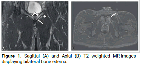
Figure 1: Sagittal (A) and Axial (B) T2 weighted MR images displaying bilateral bone edema.
All his blood investigations were within normal limits. His X-ray and MRI shows breach in anterolateral cortex with local periosteal reaction and medullary swelling (Rt.>Lt.). DEXA scan was normal. His posture and gait assessment shows:
• Increased plantar flexion of left and right ankle throughout
the gait cycle.
• Overstriding of tibia during running.
• Overpronation of foot bilaterally. Dynamic foot
assessment also shows B/L feet into overpronation [5].
Treatment
Patient first reported to us after 5 months of his injury. Initially we started with low intensity pulsed wave ultrasound therapy at the fracture site to reduce pain and start healing. After assessment we find multiple triggers in both the adductor muscles possibly because of overpronation while running. We gave injection botulinum toxin type A 10 IU in each muscular trigger in both the adductor muscles. A total of 8 sites were injected (5 on right and 3 on left). Soon after injection his pain decreased and his walking improved. To prevent his overpronation we advised him B/L insoles with medial arch supports (1 cm) and lateral wedge (6 mm). After 5 days of pain free walking, we started short distance running with icing during immediate post-activity period [6].
His physical activity was increased gradually and he didn’t complain to any pain and swelling during the next 5 weeks. After 6 weeks his X-ray was repeated which shows substantial healing. After 2 months of regular observation patient started his rigorous training and didn’t complain on any pain and difficulty. He had some dilated tortuous vein on B/L shin for which he was advised compression stockings [7].
During his follow up visit after 3 months, he is doing very well, with no stiffness and pain at the fracture site. Simultaneous regular exercises and biomechanical correction will prevent him from any further injury.
Pes planus may occur in up to 20% of the adult population, although the majority of patients are asymptomatic and require no treatment [8]. Approximately 10% (range 7%-15%) of the population with developmental flatfoot go on to develop symptoms requiring medical attention. There is some evidence to suggest that flatfeet increase the risk of stress fracture.
Our adductors are most commonly known as our inner thigh muscles. Tight adductors will make our back, our hips and even our knees hurt. For a majority of people, our adductors are directly tied to our core strength. This means if we have weak or tight adductors, our other core muscles like our abdominals are less likely to be able to function correctly. This means that if we’re struggling with back pain or a weak core, our tight adductors might be to blame [9].
An anterior tibia stress fracture is considered a high-risk stress fracture due to the combination of lack of adequate blood supply and high stress to the bone. Excessive or repetitive load on a bone can cause a mismatch between bone resorption and formation. The stress across bone will increase osteoclastic activity over osteoblastic bone formation. This mismatch causes microfracture formation when the stress to bone continues and the osteoclastic activity remains dominate. Stress fractures of the tibia can occur at any location along the shaft, but are most commonly encountered at the posteromedial cortex. Less commonly seen are stress fracture of the anterior tibia shaft. The incidence of tibial stress fractures in runners is 10%-20%.
Anterior tibial stress fractures are at high risk of delayed and nonunion due to blood supply. In a study of 50 patients with anterior tibial stress fractures, only 40% were able to return to activity and avoid surgery. The time frame for recovery could take up to a year from an anterior tibia stress fracture [10].
As intensity of training is increasing with lack of proper knowledge in managing a stress fracture is hampering and destroying the carrier of a budding sports person.
Most of the time problem is biomechanical in nature, need simple modification, if not managed proper can destroy the carrier of a budding sports person. That’s why biomechanical assessment and correction is of prime importance in any injury management.
[Google Scholar] [PubMed]
[Crossref] [Google Scholar] [PubMed]
[Crossref] [Google Scholar] [PubMed]
[Crossref] [Google Scholar] [PubMed]
[Crossref] [Google Scholar] [PubMed]
[Crossref] [Google Scholar] [PubMed]
[Google Scholar] [PubMed]
Citation: Singh S. "Treating a Case of Stress Fracture B/L Tibia with Adductor Muscle Release and Biomechanical Correction". Med Rep Case Stud, 2025, 8(6), 1-2.
Received: 18-Nov-2024, Manuscript No. MRCS-24-152805; Editor assigned: 21-Nov-2024, Pre QC No. MRCS-24-152805 (PQ); Reviewed: 05-Dec-2024, QC No. MRCS-24-152805; Revised: 01-Jan-2025, Manuscript No. MRCS-24-152805 (R); Published: 29-Jan-2025, DOI: 10.37532/mrcs.23.8.6.001-002
Copyright: © 2025 Singh S. This is an open-access article distributed under the terms of the Creative Commons Attribution License, which permits unrestricted use, distribution and reproduction in any medium, provided the original author and source are credited.