Case Report - (2021) Volume 7, Issue 10
Intravascular papillary endothelial hyperplasia or Masson's tumor is a rare benign vascular pathology. It mainly affects lower limbs and is not related to the gender or age of the patients. Evaluation by clinical history and imaging exams can be confused with other soft tissue tumors. The purpose of this article is to describe a case attended at a university hospital in Porto Alegre, Brazil. In the present study, the authors report a case of a 54-year-old male patient with the presence of nodulation on the back of the right foot. After inconclusive biopsy, the treatment of choice was surgical resection of the tumor with free margins. Anatomopathological study of the surgical specimen was compatible with Masson's tumor. A 6-month follow-up did not show any recurrence of the lesion. With this case, the authors wish to highlight this pathology among the differential diagnoses for soft tissue tumors.
Masson’s tumor • Intravascular Papillary endothelial hyperplasia • Vascular tumors
An intravascular papillary endothelial hyperplasia or Masson's tumor is an uncommon benign vascular pathology. Since 1923, when Pierre Masson first described this tumor, several articles have tried to define its origin. Although several cases are related to pre-existing vascular injuries or a history of trauma, their etiology is unknown. Assessing the clinical history and imaging tests, it is difficult to differentiate it from malignant neoplasms, especially angiosarcoma. In most cases, complete resection of the lesion is necessary, and provides treatment and definitive diagnosis of Masson's tumor.
The aim of this article is to describe a case of intravascular papillary endothelial hyperplasia treated at a university hospital in Porto Alegre and highlight the disease as a differential diagnosis for skin and subcutaneous tissue tumors that, clinically, may present similarly to malignant tumors.
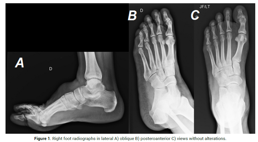
A healthy 54-year-old male patient was referred to our service complaining of nodulation on the back of the right foot 1 month ago. During this period, he did not notice mass growth. On physical examination, there was a nodulation on the back of the right midfoot of about 3 cm in its largest diameter, not hardened and mobile. Radiographs of the right foot showed that there was no bone involvement (Figure 1).

Figure 1: Right foot radiographs in lateral A) oblique B) posteroanterior C) views without alterations.
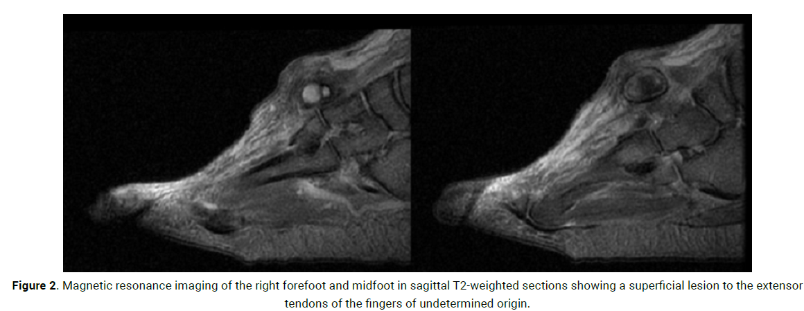
Magnetic resonance imaging of the right foot and ankle showed a lesion with heterogeneous signal located superficially to the extensor tendons of the fingers, close to the subcutaneous cellular tissue of the dorsal surface, measuring 2.6 x 2.1 x 1.5 cm, determining dorsal skin bulging, associating if swelling in adjacent soft tissues. This lesion presented hyper signal on T1 and T2, which could be related to hematic content, mild peripheral contrast enhancement and a close relationship with the path of the dorsal artery of the foot, with an indeterminate evolutionary nature and potential (Figure 2). Other aspects presented without changes.

Figure 2: Magnetic resonance imaging of the right forefoot and midfoot in sagittal T2-weighted sections showing a superficial lesion to the extensor tendons of the fingers of undetermined origin.
An open biopsy of the lesion was performed, with inconclusive anatomopathological examination, suggesting a hematoma. Then, surgical resection of the tumor was indicated. Procedure occurred after 3 months of evolution of the condition. During surgery, the tumor was found to surround the long extensor of the hallux, superficially to the extensor tendons of the toes, and it was resected, preserving the anatomy of the foot. Anatomopathological examination of the surgical specimen showed ectatic vessel with organizing thrombosis and intravascular papillary endothelial hyperplasia, compatible with Masson's tumor. The patient evolved well in the postoperative period. Six months after resection, there were no signs of tumor recurrence.
Masson's tumor is an uncommon benign vascular disease, also known as intravascular papillary endothelial hyperplasia, caused by excessive proliferation of the endothelium into normal or malformed blood vessels [1]. It affects men and women equally and can occur at any age. It comprises about 2% of vascular tumors of the skin and subcutaneous tissue [2]. This tumor can be found in any location, but mainly affects the lower limbs (about 40% of cases). It usually manifests as a palpable, painful mass of slow progressive growth [3].
Masson's tumors can be classified into 3 types. The pure or primary form is the most frequent, present in about 55% of cases, and appears in dilated vascular spaces. The second type is mixed that arises due to the presence of local alterations due to previous vascular pathology such as hemangioma, pyogenic granuloma or vascular malformation, tending to be located in intramuscular regions. The third, rare type, results from an extravascular organization secondary to a trauma-induced hematoma [4].
On imaging, it is often difficult to differentiate from malignant tumors, and final diagnosis is only possible with histopathological examination [1]. Imaging exams are especially useful to determine the extent of the lesion and its vascularity and to assist in planning surgical treatment. [3] On ultrasound, they present as hypoechoic images [5]. On Doppler, it is possible to observe the presence of central and septal vascularization [6]. During CT scan, peripheral and septal enhancement of the lesion is usually demonstrated [5]. In MRI studies, most of these lesions appear slightly heterogeneous on T1, with an isointense signal to the muscle, and may contain foci with hypersignal. On T2, they present as hyperintense masses with some foci of low signal [6]. Post-contrast, heterogeneity is marked on T1, a finding also found in malignant lesions [7].
Histopathologically, macroscopy usually presents as a well-circumscribed reddish vascularized mass with a friable surface, which may contain calcifications [3]. On microscopic study, it is possible to observe the formation of papillary structures covered by endothelial cells in the dilated vascular spaces with absence of atypia, mitotic activity, or necrosis. Thrombi can also be found within these lesions in varying sizes and different stages of organization.
Healing is achieved with total resection of the lesion with free margins. The prognosis is excellent, with no reports in the literature of local invasion or metastases in patients followed up in the long term [2]. Follow-up is necessary, as there is a low risk of lesion recurrence even with complete removal of the tumor [8]. Recurrence is most associated with cases in which the tumor has developed into pre-existing vascular lesions (the mixed types) and/or when the lesion is not completely resected [9].
We report a case of Masson's tumor followed up in our service that presented a clinical history and imaging exams that were very characteristic of the disease, but there was no possibility of excluding malignant pathologies without an anatomopathological study. Resection with free margins allowed treatment and diagnosis and it will be necessary to maintain long-term follow-up to assess tumor recurrence. Although uncommon, Masson's tumor should be considered in the differential diagnosis of malignant soft tissue pathologies in order to avoid unnecessary aggressive treatments and add morbidity to patients' lives.
T We thank the patient for allowing the case description.
The authors declare that they have no conflicts of interest.
Citation: Burguêz D. Masson’s Tumor-Case Report. Oncol Cancer Case Rep. 2021, 07 (10), 001-002.
Received: 06-Oct-2021 Published: 27-Oct-2021
Copyright: © 2021 Burguêz D, et al. This is an open-access article distributed under the terms of the Creative Commons Attribution License, which permits unrestricted use, distribution, and reproduction in any medium, provided the original author and source are credited.