
Image - (2016) Volume 5, Issue 3
A 48 year old male without any previous medical history had abdominal discomfort. On ultrasonography (US) and contrast enhanced computed tomography (CT), intra-abdominal mass located in between liver and stomach was founded. Tumour markers including AFP, CEA, CA 19-9, CA 125, and NSE were within normal range. To figure out regional relationship between mass and adjacent structures, endoscopic ultrasonography (EUS) and magnetic resonance imaging (MRI) were performed. EUS revealed a mass separated from serosa of stomach, suspicious for gastrointestinal stromal tumour (GIST). MRI revealed a lobulated contour calcification containing solid mass, suspicious for carcinoid, hemangioma, or GIST.
A 48 year old male without any previous medical history had abdominal discomfort. On ultrasonography (US) and contrast enhanced computed tomography (CT), intra-abdominal mass located in between liver and stomach was founded. Tumour markers including AFP, CEA, CA 19-9, CA 125, and NSE were within normal range. To figure out regional relationship between mass and adjacent structures, endoscopic ultrasonography (EUS) and magnetic resonance imaging (MRI) were performed. EUS revealed a mass separated from serosa of stomach, suspicious for gastrointestinal stromal tumour (GIST). MRI revealed a lobulated contour calcification containing solid mass, suspicious for carcinoid, hemangioma, or GIST.
The patient underwent mass excision. The specimen measured 12.0 × 9.0 × 7.5 cm and weighted 365 gm. On section, it showed a well circumscribed lobulated and calcified mass, measured 7.5 × 6.0 × 5.1 cm. Pathologic type was Castleman lymphadenopathy, hyaline vascular type. Postoperative course was uneventful and the patient was discharged on postoperative day 7 see Figures 1-5.
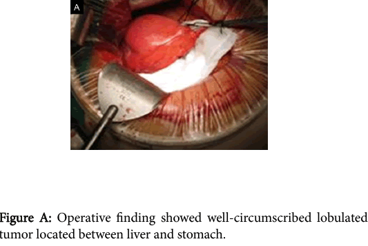
Figure 1: Operative finding showed well-circumscribed lobulated tumor located between liver and stomach.
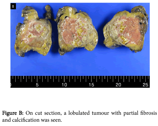
Figure 2: On cut section, a lobulated tumour with partial fibrosis and calcification was seen.
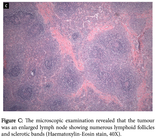
Figure 3: The microscopic examination revealed that the tumour was an enlarged lymph node showing numerous lymphoid follicles and sclerotic bands (Haematoxylin-Eosin stain, 40X).
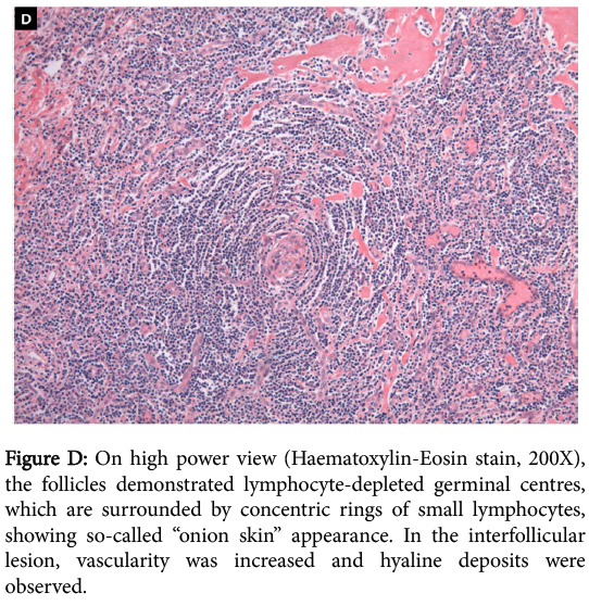
Figure 4: On high power view (Haematoxylin-Eosin stain, 200X), the follicles demonstrated lymphocyte-depleted germinal centres, which are surrounded by concentric rings of small lymphocytes, showing so-called “onion skin” appearance. In the interfollicular lesion, vascularity was increased and hyaline deposits were observed.
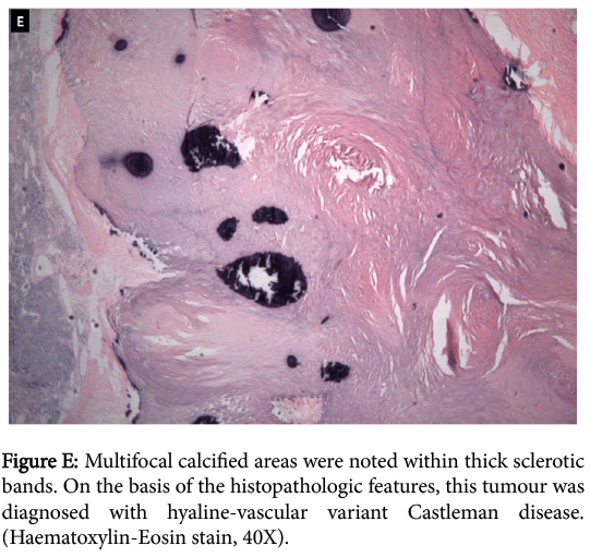
Figure 5: Multifocal calcified areas were noted within thick sclerotic bands. On the basis of the histopathologic features, this tumour was diagnosed with hyaline-vascular variant Castleman disease. (Haematoxylin-Eosin stain, 40X).
