Case Report - (2021) Volume 7, Issue 7
Wilm’s tumour is most common malignant renal tumour of childhood. Rarely, it is seen in the adults. We report a 70 year old female presenting with flank pain. Abdominal CT scan revealed a right renal mass and a clinical diagnosis of renal cell carcinoma was made. Nephrectomy was performed and a final diagnosis came as adult Wilms’ tumour. Adult Wilms’ tumour is aggressive and have poor prognosis and should be treated with the similar protocols as those used in children.
Adult wilm’s tumour, kidney, Radiation.
Wilm’s tumour or nephroblastoma, is a malignant renal tumor that arises from abnormal proliferation of metanephric blastema without differentiation into glomeruli and tubules. More than 90% of all Wilms’ tumour cases occur before age 7 with peak incidence between age 3 and 4 [1].
Mrs. XYZ, 70 year old female with previous history of cancer Breast post-operative and post chemotherapy on hormonal therapy with Tab. Tamoxifen with known diabetic and hypertensive since 6 yrs on regular medication presented to us with pain in the right loin pain for 1 month, investigated at our hospital.
USG: Right kidney 6.1 × 6.2 focal lesion with heterogenous echos in the mid and lower pole.
Chest x-ray: Normal
Complete blood picture: Within normal limits
Biochemistry: With Normal Limits
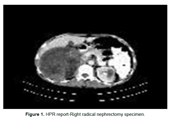
CT scan abdomen: Right kidney enlarged in size, normal in location and shows a well-defined heterogeneously enhancing mass lesion with few non enhancing necrotic areas approximately measuring 9.5 × 9.5 × 9 cm involving the lower half of the kidney, the mass is seen to displace the adjacent bowel loops ,however the fat planes are ill maintained medially there is a mass effect on the IVC with illdefined fat planes between them. Thickening of Geroto’s fascia [2] and lateral conal fascia, few perilesional lymphnodes measuring 1.2 × 0.7 cm (Figure 1).

Figure 1: HPR report-Right radical nephrectomy specimen.
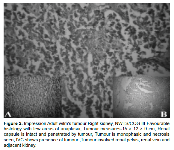
Gross & Microscopic specimen: Received Right radical nephrectomy specimen measuring 15 × 12 × 9 cm. The kidney is enlarged cut section shows a large tumour replacing almost the entire kidney. The tumour has a variegated appearance with grey white and light brown hemorrhagic and necrotic friable areas. The tumour is involving the renal sinus and renal vein grossly. The capsule is thinned out and infiltrated. The adjacent kidney is unremarkable. The ureter 8 cm in length and is unremarkable. Distal ureter 7.5 cm away from the tumour (Figure 2).

Figure 2: Impression Adult wilm’s tumour Right kidney, NWTS/COG III-Favourable histology with few areas of anaplasia, Tumour measures-15 × 12 × 9 cm, Renal capsule is intact and penetrated by tumour, Tumour is monophasic and necrosis seen, IVC shows presence of tumour ,Tumour involved renal pelvis, renal vein and adjacent kidney.
Wilms’ tumour, or nephroblastoma, is most common renal tumour in children accounting for 9% of all paediatric solid tumour malignant renal tumour that arises from abnormal proliferation of metanephric blastema. There is no significant difference between the radiological appearance of Wilms’ tumour seen in either children or adults. About 75-80% of the cases have similar clinical and radiological findings.
Adult Wilms’ tumour is diagnosed based on the criteria given by Kilton, Mathews and Cohen [3]. These include:
• The tumour under consideration should be a primary renal neoplasm.
• Presence of primitive blastemic spindle or round cell component.
• Formation of abortive or embryonal tubules or glomerular structures.
• No area of tumour diagnostic of renal cell carcinoma.
• Pictorial confirmation of histology and
• Patient’s age >15 years.
Differential diagnosis of an adult Wilms’ tumour
• Primary renal cell carcinoma
• Lymphoma
• Rhabdomyosarcoma
• Metastatic small cell tumours from lung
About half of the patients have stage 3 or 4 disease [4]. The most frequent places of metastasis are lung, liver, bones, skin, brain and contralateral kidney. Metastasis rates for children and adults are 10% and 29%, respectively [1]. The cytogenetic changes of isochromosome 17 q are frequently seen with these types of tumour in adult age group [5]. There is no standard therapy for treating patients with adult Wilms’ tumour due to the rarity of this subset and paucity of literature available. The National Wilms’ Tumour Study-3 (NWTS) showed that for low risk patients (favorable histology [FH], Stage I/II), less aggressive therapies were not demonstrably worse than more intensive procedure [6] however results of treatment for adult patients classified with Stage I/II disease published more recently are promising, with an outlook similar to that for children. An update from the NWTS group about treatment outcomes in adults with favourable histology Wilms’ tumour (FHWT) described 45 patients treated in the modern era. The overall survival rate was 82%.
Treatment should consist of multimodal therapy with surgery, chemotherapy (dactinomycin plus vincristine plus doxorubicin) for 15 months and tumour- bed irradiation with IMRT technique to a dose of 1980 cGy/11# to the flank keeping the normal tissue dose within limits and this was according to the NWTS and the experiences associated with adult Stage III disease [7]. Prognosis for adult patients with unfavorable histology and Stage IV disease (hematogenous metastases) is poor despite aggressive multimodal therapy. Furthermore, for patients with recurrent disease, encouraging results have been reported. Recently long-term remissions have been achieved using high dose chemotherapy, radiotherapy and allogeneic bone marrow transplantation or combination chemotherapy with cisplatin and etoposide [8-10].
Nephrectomy was performed and a final diagnosis came as adult Wilms’ tumour. Adult Wilms’ tumour is aggressive and have poor prognosis and should be treated with the similar protocols as those used in children.
Citation: Raghavendra D Sagar. Adult Wilm’s Tumour- A Rare Tumour. Oncol Cancer Case Rep. 2021, 07 (7), 001-002.
Received: 16-Jun-2021 Published: 23-Jul-2021
Copyright: © 2021 Sagar RD. This is an open-access article distributed under the terms of the Creative Commons Attribution License, which permits unrestricted use, distribution, and reproduction in any medium, provided the original author and source are credited.